MRI and blood biomarker analysis reveals what happens to the brain after soccer heading
New research conducted at NeuRA identified microstructural, functional and chemical changes after controlled soccer heading.
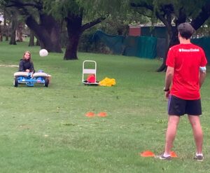 This is the first randomised controlled trial to use MRI to examine how the brain responds to soccer heading. Conducted in a highly controlled environment, adult soccer players headed a regulation soccer ball 20 times in 20 minutes, with balls launched at a constant speed from a machine.
The soccer players also completed a separate control condition, whereby they kicked the ball, allowing the researchers to isolate the effects of heading. More detail about the background and methods can be found in our previous NeuRA Imaging blog post: https://imaging.neura.edu.au/research-spotlight-biomarkers-of-subconcussive-impacts-in-sport-the-bump-trial/.
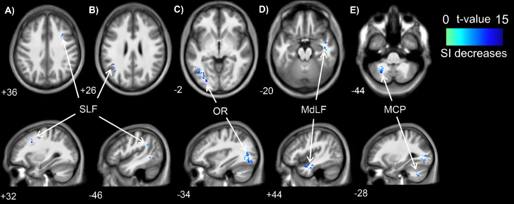
Using five high-quality advanced MRI acquisitions approximately 45 minutes after heading, the researchers detected two subtle changes. They found reductions in tissue conductivity, via the sequence known as electrical properties tomography, in several predominantly posterior white-matter brain regions (figure below).
The largest cluster was found in the optic radiation, which transmits visual information to the visual cortex. This is interesting given the well-established link between more severe head impacts (e.g., concussion) and visual disturbances.
Locations of significant clusters from electrical properties tomography data (above) and statistical details (below) between conditions. Cooler (blue) colours represent higher t values and a decrease in signal intensity. Location of each sagittal and axial slice in Montreal Neurological Institute space are indicated at the bottom left of each slice. Abbreviations: MCP–middle cerebellar peduncle; MdLF–middle longitudinal fasciculus; MNI–Montreal Neurological Institute; OR–optic radiation; SI–signal intensity; SLF–superior longitudinal fasciculus.
The second was elevated metabolite levels of total N-acetyl aspartate and creatine in the primary motor cortex, via the sequence proton magnetic resonance spectroscopy. This may signify elevated brain metabolism in this region.
In addition to the MRI findings, highly sensitive blood analysis techniques identified an increase in proteins GFAP and Nf-L in peripheral blood. These proteins are expressed in glia and axons of neurons in the brain, and an increase represents disruption to these brain cells. All these changes were observed in the absence of detectable alterations in cognitive function, and few symptoms.
The researchers note that more research is needed to determine the clinical significance of these changes, but nonetheless, they provide a glimpse into the otherwise covert responses of the brain to heading. In the meantime, they suggest footballers should consider whether excessive heading is necessary, particularly during training sessions where the amount can be limited.
Research: Nathan Delang, et al., ‘The acute effects of non-concussive head impacts on brain microstructure, chemistry and function in male soccer players: A pilot randomised controlled trial’. (Sports Medicine – Open) DOI:
Funding: This study was primarily supported by funding from the Lambert Initiative for Cannabinoid Therapeutics (a philanthropically funded centre for medicinal cannabis research at the University of Sydney). The philanthropic donor did not have any input into the investigation. Additional funding support was provided internally from Griffith University and Monash University.
Read the full article
Delang, N., Robertson, R.V., Tinoco Mendoza, F.A. et al. The Acute Effects of Non-concussive Head Impacts on Brain Microstructure, Chemistry and Function in Male Soccer Players: A Pilot Randomised Controlled Trial. Sports Med – Open 11, 77 (2025). https://doi.org/10.1186/s40798-025-00867-0
Recent Comments