New Insights Into Chiari: What Happens During a Cough?
A team of researchers at NeuRA Imaging has captured the most detailed MRI footage to date of what happens inside the head and neck during a cough in people with Chiari malformation, challenging decades of assumptions about how the condition causes sudden, debilitating headaches.
For years, clinicians have believed that cough‑triggered headaches in Chiari malformation stem from the cerebellar tonsils obstructing the flow of cerebrospinal fluid (CSF) at the foramen magnum. But what the NeuRA team observed tells a very different story.
In the study recently published in Computers in Biology and Medicine, researchers, including Dr Rob Lloyd and Professor Lynne Bilston used a combination of high‑resolution anatomical imaging and fast, ungated phase‑contrast MRI on NeuRA Imaging’s Philips Ingenia 3T CX MRI scanner, to watch CSF accelerate toward the brain with every cough.
To their surprise, the fluid never stopped flowing, even in patients with severe tonsillar herniation. Lateral channels remained open, and the foramen magnum acted less like a blocked tunnel and more like a crowded roundabout that still keeps traffic moving.
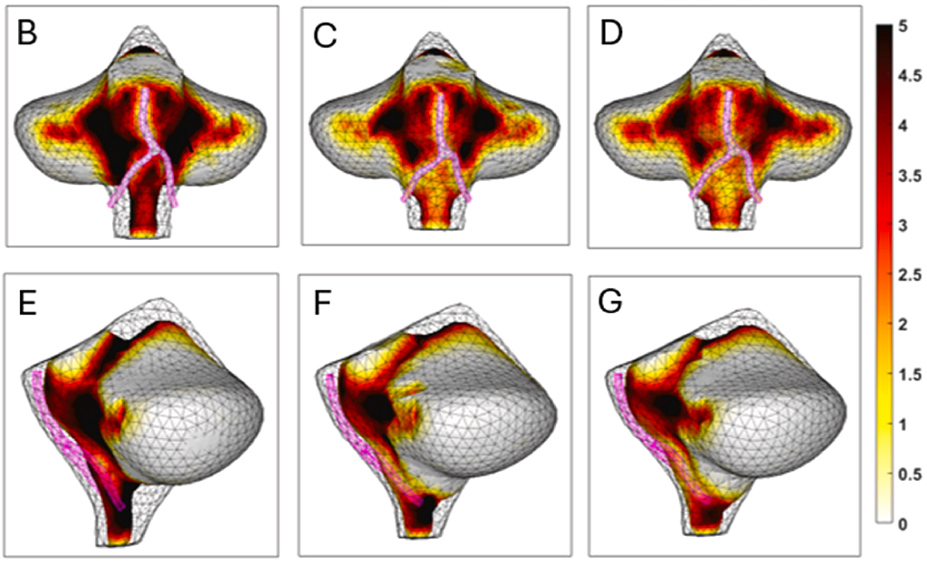
What did change was speed. During the explosive phase of the cough, CSF velocities in Chiari patients spiked to more than twice those seen in healthy volunteers. The faster the fluid moved, the more force it exerted on the tightly packed structures at the craniocervical junction.
And in those who suffer cough‑associated headaches, one anatomical feature stood out. The researchers discovered a narrowed CSF space at the front of the brainstem, particularly around the vertebral and basilar arteries. This tight anatomical corridor means that when CSF accelerates during a cough, it has less room to move, potentially generating stress on nearby vessels and cranial nerves.
Lead author Dr Rob Lloyd, from Neuroscience Research Australia and UNSW Sydney, says the findings could reshape our understanding of Chiari‑related pain.
“Our study shows that the obstructing tonsil may be a red herring, and that the main issue is likely CSF being forced over sensitive tissues in front of the brain stem. If we can confirm this, it will provide a reliable clinical marker that could help standardise the treatment of patients with Chiari malformation.
Dr Rob Lloyd, School of Biomedical Engineering UNSW, and Neuroscience Research Australia”
The study also suggests why surgery doesn’t always relieve symptoms. Standard decompression focuses on enlarging the posterior fossa behind the cerebellum. But the team’s shape‑analysis models reveal that the critical pressure changes during a cough may actually occur in the anterior CSF space, where most surgeries do not directly intervene.
The deep learning models used to automate the analysis of the imaging used in this study is available via the Open Science Framework https://osf.io/hte5y/
The work highlights the research value of modern MRI systems capable of real‑time imaging. The Philips Ingenia 3T CX at NeuRA Imaging allowed researchers to visualise rapid CSF motion across multiple planes, something earlier pencil‑beam MRI methods could not achieve.
For patients, the message is cautiously hopeful. The findings show that symptoms may not be due to an obstructed brain, as once feared, but rather to complex fluid dynamics interacting with individual anatomy. That opens the door for new diagnostic markers, new surgical targets and, potentially, better prediction of which patients will benefit most from treatment.
The full research publication is available here via Science Direct.
Recent Comments