A glimpse into brainstem functional oscillations across individual migraine cycles
Recently published in NeuroImage: Clinical is a paper from the PhD thesis of Noemi Meylakh, using the Philips 3T TX MRI scanner, to measure resting functional oscillations in controls and episodic migraineurs across the span of 30 days (doi: 10.1016/j.nicl.2021.102630).
Since the 17th Century, philosophers have struggled with understanding the pathophysiology of the ever-elusive migraine disorder. Wolff’s “vascular theory” held true for over four decades.
However, vasodilation and constriction of blood vessels could not explain for all aspects of migraine.
And so, the understanding of migraine pathophysiology shifted from purely a disorder of blood vessels to a “highly choreographed interaction between major inputs from both the peripheral and central nervous systems” (Pietrobon & Moskowitz, 2013).
Whilst there has been a considerable amount of investigation into neural mechanisms involved in the ictal (during a migraine) and interictal (pain-free) phases of migraine, exploration of the premonitory (24-hour period preceding a migraine headache) has been more difficult.
Despite this, several investigations including our own, have shown for the first time that functional activity of pain-modulatory regions of the brainstem, and the homeostatic centre, i.e. the hypothalamus, have a sudden spike in activity in this critical phase.
Furthermore, this spike in activity is cyclic, normalising after the resolution of the headache, i.e. in the postictal phase. Understanding sudden neural changes in activity in this phase is imperative in unravelling the nature of the processes that take place during the initial stages of a migraine event.
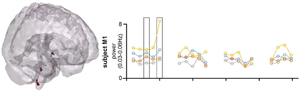
The best way to track neural events that take place in the leadup to a migraine attack is to assess brain function each day in a single individual. In this paper, we performed MRI scans in three migraineurs and five healthy controls at the same time five days a week for four weeks.
The aim of the study was to corroborate the findings from premonitory studies, in other words, to see if function of brainstem pain-modulatory sites oscillates over the migraine cycle, with a spike in activity 24-hours prior to a migraine headache. Indeed, this is exactly what we found.
We measured resting functional MRI and found that functional activity in the premonitory phase of individual migraine cycles was associated with an increase in activity in important pain-modulatory regions.
These findings significantly contribute to our understanding of the migraine cycle, especially the underlying neural events that take place in the leadup to a migraine attack. Hopefully, studies like these can inform migraine prevention and treatment strategies.
Recent Comments