Chronic low back pain is one of the leading causes of disability worldwide, and it becomes more common as we age. In Australia, around one in six adults report back problems. For older people, persistent back pain brings an extra and often overlooked risk: it is linked to poorer balance, a greater fear of falling, and more falls. We still don’t know much about why this happens, and in particular what is going on in the brain.
A new study led by Mahsa Seydi and Associate Professor Kimberley van Schooten from UNSW Sydney and Neuroscience Research Australia (NeuRA), published in the European Journal of Pain, set out to find out. The team used brain imaging carried out at NeuRA Imaging to look at how brain structure and tissue properties relate to real-world walking, physical function and thinking skills in older adults living with chronic low back pain.
Measuring how people walk in everyday life
Thirty-three adults aged 50 and over with chronic low back pain took part. Instead of relying only on clinic-based tests, each participant wore a small waist-worn sensor for a full week as they went about their normal lives. The sensor recorded how smoothly and steadily they walked. This measure of “daily-life gait quality” has previously been shown to predict future falls.
Participants also completed tests of physical function (walking speed, standing balance and rising from a chair), reported their physical activity, and did quick tests of thinking skills. One of these was the ReacStick, a “ruler drop” task that tests how fast someone can stop themselves from reacting. That kind of rapid braking is important for recovering your balance.
Three ways of looking at the brain
Each participant had an MRI scan on NeuRA Imaging’s Philips 3T scanner, using three complementary approaches:
- Structural MRI to measure the size of different brain regions
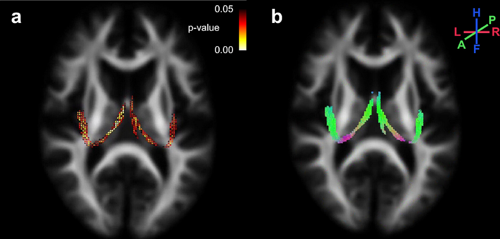
- Diffusion MRI to look at the brain’s white matter “wiring” that connects regions
- Magnetic Resonance Electrical Properties Tomography (MREPT), an emerging technique that maps how well brain tissue conducts electricity, which may reflect subtle changes at a microscopic level
Two axial brain MRI images. The left image shows highlighted white matter regions in yellow and red where fibre measures were linked to physical performance. The right image shows the same regions as green fibre tracts running front to back through the brain.
What did they find?
Everyday walking quality was not linked to brain size or white matter wiring. It was linked to tissue conductivity in the cerebellum, pallidum and thalamus. These deep brain regions are known to be important for coordination and controlling movement. This suggests that subtle changes in brain tissue may affect balance before any visible structural change can be seen on a standard scan.
Other measures showed different patterns:
- Physical function was related to the size of deep brain regions such as the hippocampus, thalamus and putamen, and to white matter pathways that carry visual information. This may mean that people with back pain rely more on vision to keep their balance.
- Physical activity was linked to conductivity in regions involved in movement, navigation and motivation.
- Rapid inhibitory control (stopping a reaction quickly) was linked to widespread movement-related white matter pathways and to conductivity in the orbitofrontal cortex, a frontal brain region involved in self-control.
Why it matters
The findings suggest that balance and fall risk in older people with chronic back pain involve the brain as well as the back. Movement, thinking and activity all draw on overlapping brain networks. This supports a broader approach to care that could include balance and coordination training and combined cognitive-motor training, alongside conventional musculoskeletal rehabilitation.
The authors note that this was a small, cross-sectional pilot study with relatively healthy and active participants and no pain-free comparison group. The results should be seen as a first step, and they open the door to larger and longer-term studies. The study also shows that MREPT could be a useful addition to the neuroimaging toolkit, since it picked up brain–behaviour relationships that conventional MRI measures did not.
Read the full study
For researchers interested in the multimodal imaging methods, the fixel-based diffusion analysis, or the use of MREPT in pain and mobility research, the full open access article is available in the European Journal of Pain:
Seydi, M., Delbaere, K., Ambrens, M., Richardson, J. K., Rae, C. D., Cao, J., Sachdev, P. S., Lavrencic, L., Green, M. A., & van Schooten, K. S. (2026). Neural Correlates of Fall Risk in People Aged 50 and Over With Chronic Low Back Pain: A Multimodal MRI Study. European Journal of Pain, 30, e70380. https://doi.org/10.1002/ejp.70380
Recent Comments