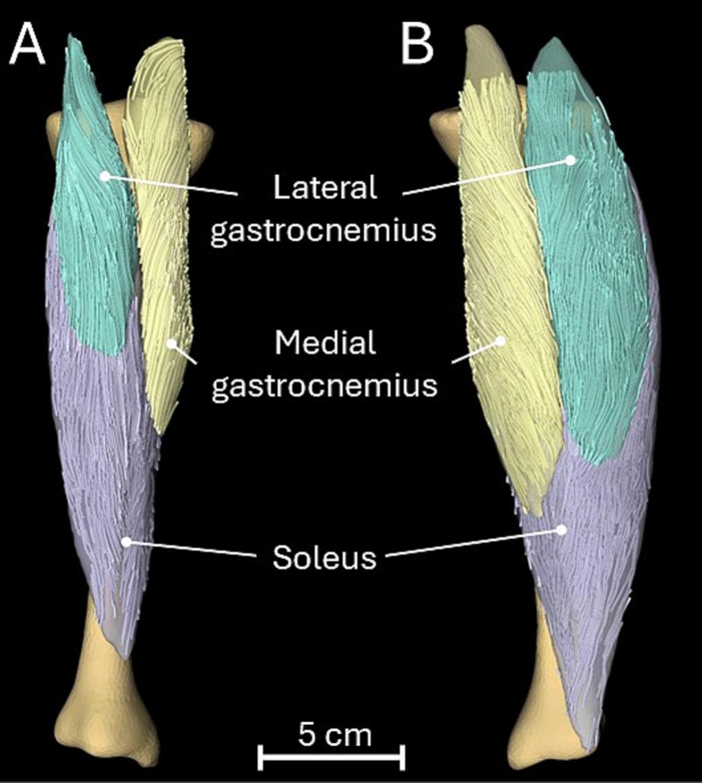
New imaging research reveals that children with cerebral palsy don’t lose calf muscle size in one uniform way. Instead, each muscle sacrifices either fibre length or fibre width to protect its own specialised job, whether that’s generating force or allowing a full range of motion.
by Bart Bolsterlee
Cerebral palsy (CP) is the leading cause of physical disability in children worldwide. Previous research has shown that most muscles in most children with CP are substantially smaller than those in age- and sex-matched typically developing children (Bolsterlee et al., 2025, PNAS). Small muscles contribute to the loss of strength and mobility many children with CP live with.
However, while muscle size is an important predictor of muscle strength, the lengths and arrangement of muscle fibres within the muscle belly play a more important role in a muscle’s ability to generate force and movement.
 Muscle fibres are typically several centimeters long but only 10 to 100 micrometer in diameter – think of it as the size of a hair. When children’s muscles grow, these fibres can get longer, thicker, or both. Longer fascicles let a muscle stretch and shorten over a wider range, which matters for joint range of motion. Wider fibres (increases in cross-sectional areas) result in increased strength.
Using advanced protocols and expertise available at NeuRA Imaging’s 3T Philips Ingenia CX MRI scanner, including diffusion tensor imaging, we measured the lengths and cross-sectional areas of lower leg muscles of 69 children with CP and 195 typically developing children aged 5 to 18 years. Each child was tracked for up to 3 years.
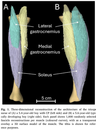
We found that the three major calf muscles, the soleus and the medial and lateral gastrocnemius, vary in how they adapt in children with CP. While in all muscles the fibres were usually narrower (smaller cross-sectional areas) and shorter compared to typically developing children, the degree of these adaptations varied strongly between muscles.
In the soleus of children with CP, the cross-sectional areas of the fibres were relatively preserved (~6% smaller than in age- and sex-matched typically developing children) but the fibres were substantially shorter (by ~25%). In the medial gastrocnemius, fibre lengths were relatively preserved (~7% shorter) but cross-sectional areas were substantially reduced (by ~28%). In the lateral gastrocnemius, the reductions in cross-sectional areas (17%) were similar to reductions in fibre lengths (15%).
These findings show that the soleus adapts primarily to preserve its force-generating capacity (at the expense of its ability to generate high forces) while the medial gastrocnemius adapts to preserve its range of motion (at the expense of loss of range of motion). Even in muscles that are closely linked in function, volumetric loss is the result of different adaptation mechanisms at the muscle-fibre level.
The findings of this large and detailed imaging study provide important information for researchers and clinicians to understand how CP impairs physical performance, and may help the development of more effective interventions.
Paper:
Bolsterlee, B., Chow, B.V.Y., Davies, S., Morgan, C., Rae, C.D., Warton, D.I., Novak, I., Lancaster, A., Popovic, G.C., Seydi, M., et al. (2026). Triceps surae muscle architecture in ambulant children with cerebral palsy: architectural adaptations vary between children and muscles. Journal of Biomechanics, 202, 113286. https://doi.org/10.1016/j.jbiomech.2026.113286
Related papers:
Chow BVY, Davies S, Morgan C, Rae CD, Warton DI, Novak I, Lancaster A, Popovic GC, Rizzo RRN, Rizzo CY, Ball IK, Herbert RD, Bolsterlee B. Architecture of lower leg muscles in children: Reference curves and potential mechanisms of growth. J Anat. 2026 Jul;249(1):54-67. doi: 10.1111/joa.70082.
B. Bolsterlee, B.V.Y. Chow, J. Yu, S. Davies, C. Morgan, C.D. Rae, D.I. Warton, I. Novak, A. Lancaster, G.C. Popovic, R.R.N. Rizzo, C.Y. Rizzo, I.K. Ball, & R.D. Herbert, Childhood muscle growth: Reference curves for lower leg muscle volumes and their clinical application in cerebral palsy, PNAS 122 (14) e2416660122, https://doi.org/10.1073/pnas.2416660122 (2025).
Recent Comments