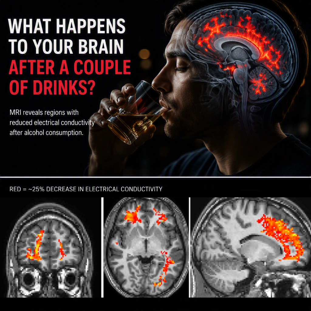
We’ve just shared one of our favourite MRI visualisations on the new NeuRA Imaging Instagram account, highlighting how advanced MRI technology can reveal changes in the brain after alcohol consumption.
Most people are familiar with the effects of alcohol, whether it’s feeling more relaxed, slightly unsteady, or finding it harder to concentrate. But what is actually happening inside the brain during this process? Using specialised MRI techniques, researchers at NeuRA Imaging investigated how alcohol alters the electrical properties of brain tissue in healthy social drinkers.
The study led by Professor Lindy Rae and Dr Jun Cao used a technique known as Magnetic Resonance Electrical Properties Tomography (MREPT), which allows researchers to estimate the electrical conductivity of tissues non-invasively. Electrical conductivity is influenced by the concentration and mobility of ions within tissue and provides a unique window into the biophysical state of the brain.
After participants consumed a dose of vodka, MRI scans revealed several regions where brain electrical conductivity decreased. In some areas, conductivity was reduced by approximately 25%, as shown by the red regions in the images below.
These findings are particularly interesting because alcohol is often associated with its behavioural effects, but much less is known about how it alters the brain’s physical and electrical environment. Alcohol interacts with a number of neurotransmitter systems, including GABA and glutamate pathways, contributing to its overall depressant effects on the central nervous system. The conductivity changes observed in this study may reflect some of these underlying physiological processes.
While alcohol can produce mild stimulant effects at very low doses, the predominant effect of larger amounts is sedation. This is why reaction times slow, coordination becomes impaired, and cognitive performance declines as blood alcohol levels rise. The MRI measurements provide evidence that these functional changes are accompanied by measurable alterations in the brain’s electrical properties.
This work also highlights the growing capabilities of modern MRI. While MRI is widely known for producing detailed anatomical images, advanced research techniques can measure much more than structure alone. Researchers can now investigate blood flow, connectivity, brain chemistry, tissue microstructure, mechanical properties, and even electrical characteristics, all without invasive procedures.
At NeuRA Imaging, our Philips Ingenia CX 3T MRI system supports a wide range of advanced neuroimaging and medical imaging applications, enabling researchers to explore questions that would have been impossible to study just a few decades ago.
📸 Follow @neuraimaging on Instagram for more neuroimaging research, MRI visualisations, behind-the-scenes content, and the latest updates from our open-access 3T MRI research facility.
📖 Read the original research paper: https://doi.org/10.1002/jmri.29548
📚 Read our more detailed article about this study: https://imaging.neura.edu.au/ethanol-reduces-brain-electrical-tissue-conductivity-in-social-drinkers-in-a-region-specific-pattern/
Recent Comments