RESEARCH SPOTLIGHT Muscle Growth in the Lower Extremity
NeuRA Imaging is providing high-quality data for a comprehensive study investigating muscle growth in children.
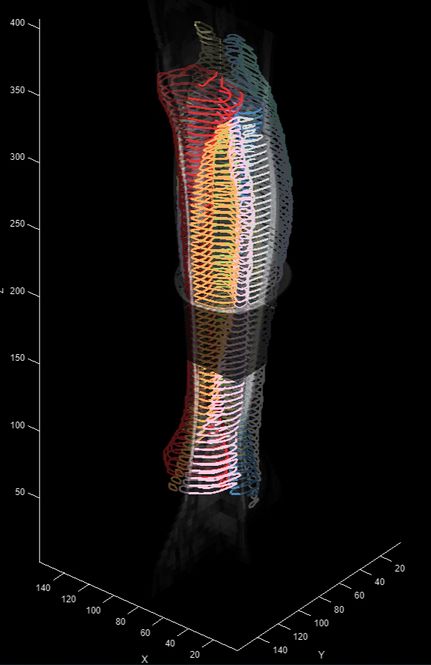
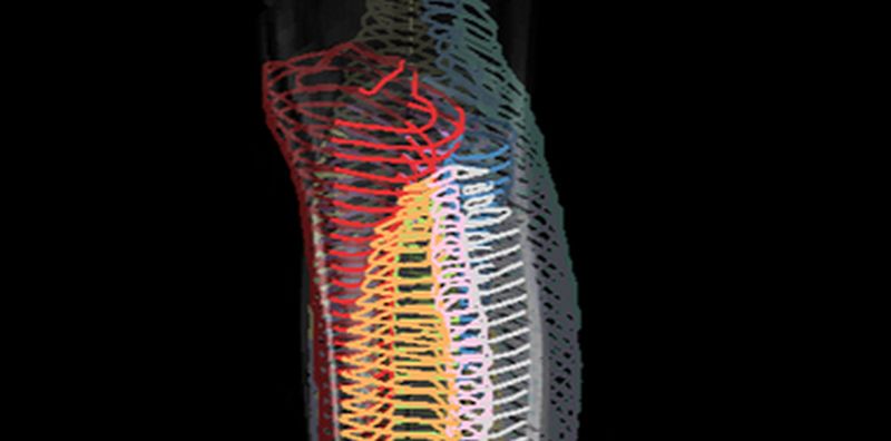
Reconstructed segmentations of muscle and bone from T1-weighted images acquired on the Philips 3T MRI at NeuRA Imaging.
A collaboration between the Cerebral Palsy Alliance Research Institute, University of New South Wales and Neuroscience Research Australia (NeuRA) is the first longitudinal study looking at muscle growth in children using magnetic resonance and diffusion tensor imaging methods.
The MUGgLE study, as it is known, is spearheaded by Rob Herbert, Bart Bolsterlee and Caroline Rae and includes the acquisition of high-quality MRI anatomical and diffusion scans of the legs in typically developing infants and children, and children with cerebral palsy.
The study aims to describe muscle growth changes in normally developing infants and children, and compare them with muscle growth observed in children with cerebral palsy.
The study is on-going and has so far acquired datasets on 57% of the targeted 320 participants with initial data analysis highlighting the performance of deep learning models for automatic segmentation of anatomical MRI scans of children with and without cerebral palsy.
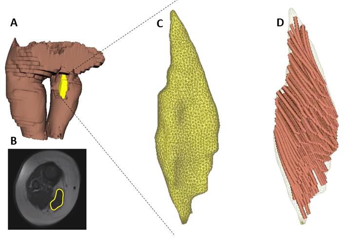
Three scans have been made of infants (3 months of age). Figure 1 demonstrates the feasibility of using MR-DTI for measuring infant muscle architecture. To a rough approximation, fascicle length, physiological cross-sectional area (PCSA) and muscle volume scaled with the first, second and third powers of tibia length, respectively. However, the generality of this scaling needs to be further tested with more data.
For more information on the MUGgLE study please visit https://muggle.neura.edu.au/
The MUGgLE study includes 3D surface reconstruction of the lower-limbs of infants and children from T1-MRI scans acquired at NeuRA Imaging.
Recent Comments