Stunning brain images acquired using high-resolution diffusion-weighted imaging
Ever wondered what Diffusion-Weighted Imaging would look like when scan time is not an issue?
A recent project led by Mark Schira and George Paxinos that constructed a histology grade atlas of the human brain based on in vivo diffusion MRI set out to answer that question.
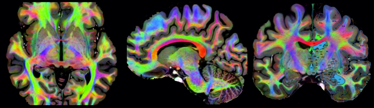
DWI images were collected with the assistance of Clinical Scientist Iain Ball on the Philips Ingenia CX 3T MRI scanner at NeuRA Imaging with acquisition parameters: 1.25mm isotropic voxels, FOV 240x200mm, 118 slices, 32 directions, 5 b-factor averages, b-val=1000, TE=60ms, TR=26.5s, SENSE=3, SPIR (Spectral Saturation with Inversion Recovery) fat saturation, fat shift direction A to P.
The results (analysed using MRTrix) are of stunning image quality and unprecedented detail. The figure above, for example, shows an acquired diffusion image overlaid with diffusion encoded colour maps with annotated anatomical landmarks.
If you want to know more about the project, visit our website here at hba.NeuRA.edu.au
Recent Comments