
Unravelling the Mystery of Sleep Apnoea: How Dynamic MRI Helps Decode Tongue Muscle Behaviour
Have you ever wondered why some people experience interrupted breathing during sleep? It turns out that the way our tongue muscles behave during sleep plays a crucial role, and researchers at NeuRA are using dynamic MRI techniques to uncover the secrets behind this phenomenon.
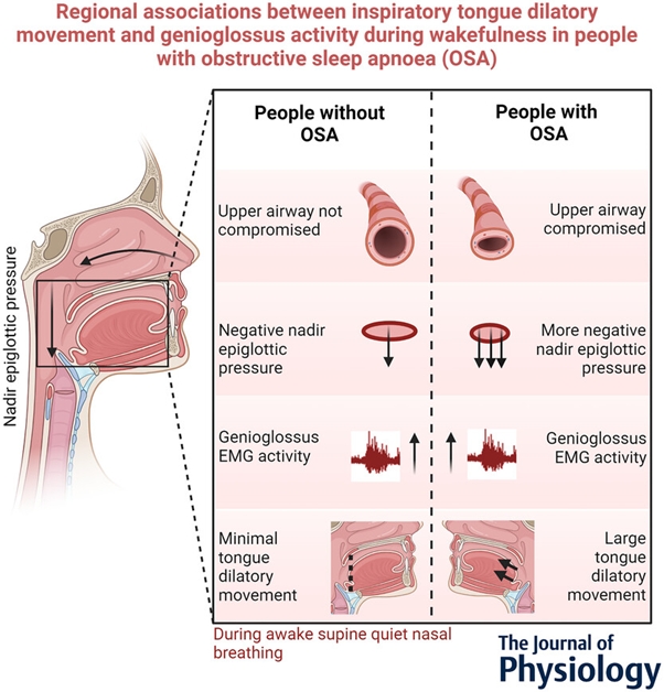
Note: Graphical abstract taken from the published article.
A recent study by a team of scientists at Neuroscience Research Australia led by Professor Lynne Bilston, investigated how the tongue moves during awake breathing in individuals with a condition called obstructive sleep apnoea (OSA). OSA is a disorder where the airway repeatedly becomes blocked during sleep, leading to pauses in breathing and disrupted sleep patterns.
The researchers were particularly interested in a muscle of the tongue called the genioglossus, which helps keep the airway open during breathing. They wanted to understand how the tiny electrical signals from the brain that cause this muscle to contract relate to the movement of the tongue during breathing and how this relationship might differ in people with OSA when compared to people without OSA.
To do this, they accessed the Philips 3T Ingenia CX scanner at NeuRA Imaging and used a technique called tagged MRI to watch the tongue in action.
The technique enables researchers can manipulate the magnetic field from the MRI to put tags on the tongue as it typically contracts and moves forward during inspiration and relaxes backward during expiration. The researchers also used electromyography (EMG) to measure electrical muscle activity during the breathing cycle.
In most cases, they observed that when the activity of the genioglossus muscle was higher, the tongue moved forward more during inspiration, which helps keep the airway open. This is expected since a larger muscle activity is often thought to result in a larger muscle contraction. However, surprisingly, in some instances, there was a disconnect between muscle activity and tongue movement, especially in people with OSA. In those cases, a larger anterior tongue movement was associated with a smaller muscle activity.
This disconnect could be one of the reasons why individuals with OSA experience airway collapse during sleep. By understanding these complex relationships between muscle activity and tongue movement, researchers hope to better understand OSA. This is important to develop new and better treatment options for patients. However for the time being, we need to find out why the electrical signal from the brain is not doing its job.
In summary, the study highlights the importance of tongue muscle function in maintaining an open airway during sleep, and how MRI is helping researchers unravel the complexities of this process. By understanding the nuances of tongue muscle behaviour, we can move closer to developing more effective treatments for sleep apnoea and ultimately improve sleep quality for millions of people worldwide.
This study was recently published in The Journal of Physiology (open access): https://physoc.onlinelibrary.wiley.com/doi/full/10.1113/JP285187 and was highlighted in a Translational Perspectives article by Professor O’Halloran, University College Cork (Ireland) https://doi.org/10.1113/JP285841.
Recent Comments