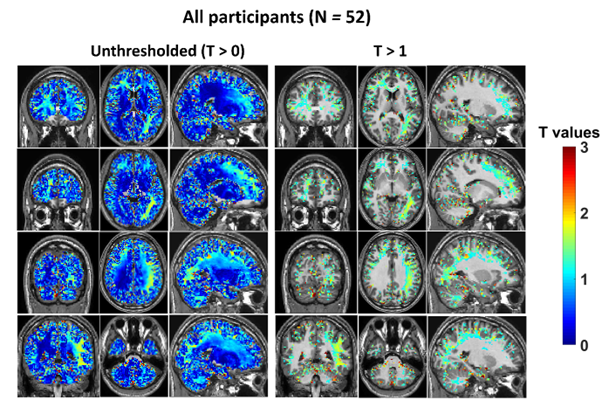
Ethanol reduces brain electrical tissue conductivity in social drinkers in a region-specific pattern
A new publication led by Lindy Rae and Jun Cao at NeuRA Imaging shines light on the effect of alcohol on brain conductivity in social drinkers. Ethanol has two main effects on the brain. It acts as a stimulant at very low concentrations but is also a sedative, substantially reducing brain metabolism, particularly through activity …
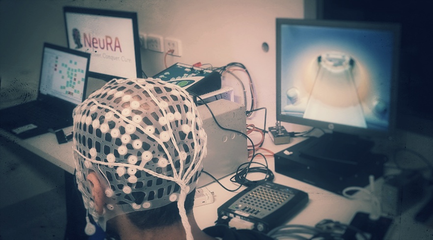
NeuRA Imaging’s newest tool enables simultaneous EEG and MRI
Neuroscience Research Australia (NeuRA) now offers researchers access to the Compumedics MicroMagLink RT System, enabling simultaneous EEG and MRI studies with optimized imaging protocols. Researchers seeking to utilize advanced imaging technology can now access the Compumedics MicroMagLink RT System at NeuRA Imaging. The system designed for integration with the 3T Philips MRI Scanner and its …

Unravelling the Mystery of Sleep Apnoea: How Dynamic MRI Helps Decode Tongue Muscle Behaviour
Have you ever wondered why some people experience interrupted breathing during sleep? It turns out that the way our tongue muscles behave during sleep plays a crucial role, and researchers at NeuRA are using dynamic MRI techniques to uncover the secrets behind this phenomenon. A recent study by a team of scientists at Neuroscience Research …
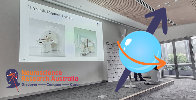
NeuRA Imaging: Advancing Knowledge in MRI Physics
NeuRA Imaging has hosted a series of educational sessions teaching the fundamentals of MRI Physics. Led by Philips Clinical Scientist, Iain Ball, the initial sessions of the course focused on the foundational aspects of MRI Physics. Topics covered included magnetism and magnetic fields, nuclear magnetic resonance, MR instrumentation, relaxation, spin density, and T1, T2, T2* …
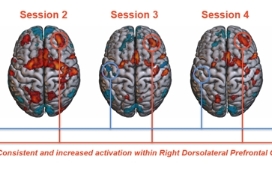
RESEARCH SPOTLIGHT: Needles in a haystack – searching for a consistent placebo analgesia responder
We know that many people can experience placebo analgesia – but are these responses stable over time? Borrowing from the designs of longitudinal preclinical investigations, researchers at NeuRA, led by Professors Luke Henderson and Kevin Keay, have recently completed preliminary data collection investigating the consistency of placebo analgesia, combining genetic, behavioural, biological, and brain imaging …
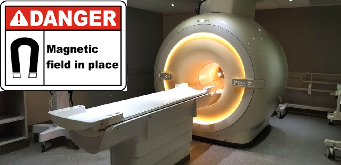
MRI Safety at NeuRA Imaging: What You Need to Know Before Using Our Philips 3T Scanner
NeuRA Imaging is committed to ensuring the highest standards of MRI safety, offering an induction course for researchers who wish to use our state-of-the-art MRI scanner. Our Philips 3T MRI scanner isn’t just a top-of-the-line research tool; it’s also an open-access facility fully dedicated to academic, industry, and clinical research. But we offer more than …
Recent Comments