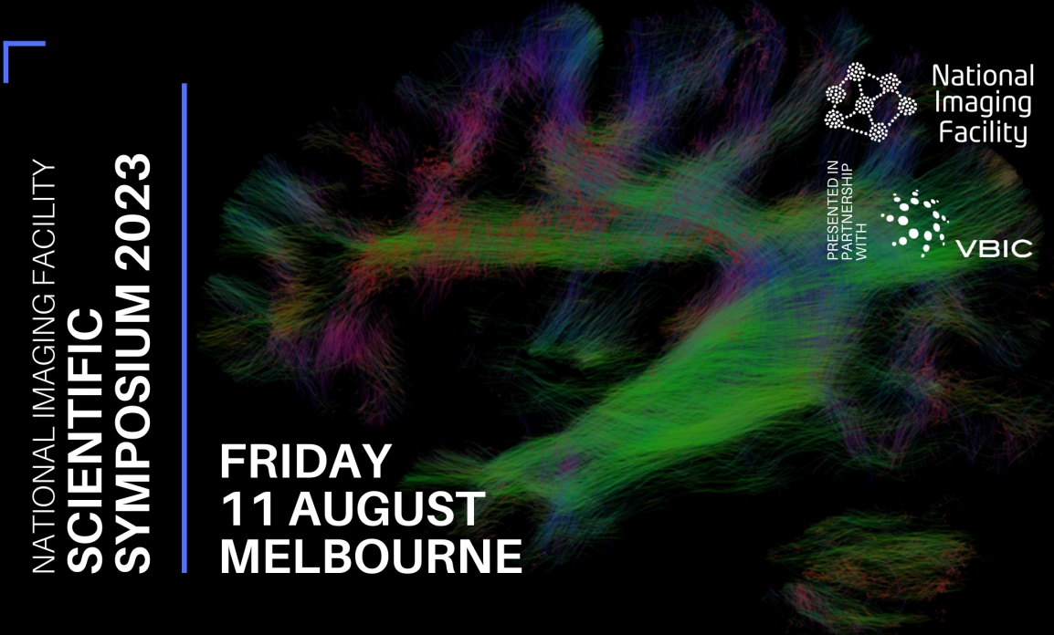
National Imaging Facility Annual Scientific Meeting 2023
NeuRA Imaging is an integral member of The Australian National Imaging Facility and we invite you to participate in our annual scientific symposium. YOU’RE INVITED: National Imaging Facility Scientific Symposium 2023 The 2023 National Imaging Facility Scientific Symposium is presented in partnership with the Victorian Biomedical Imaging Capability (VBIC). Friday 11 August 2023 8:30am – …
RESEARCH SPOTLIGHT: Using Magnetic Resonance Elastography (MRE) To Define The Shear Properties Of Fat Under Load
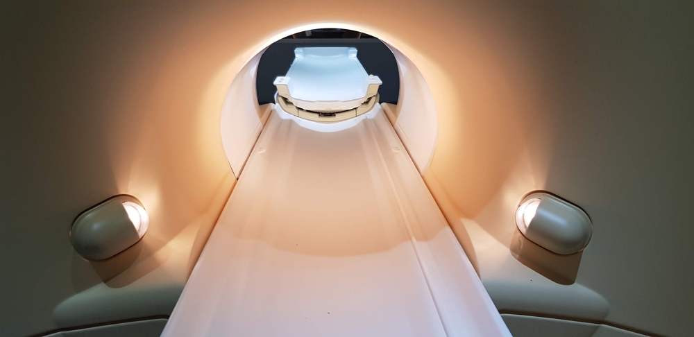
We know that you have heard of MRI, but have you heard of MRE? Magnetic resonance elastography, or MRE as it is commonly known as, is an emerging MR technique that allows the stiffness of internal tissues to be measured quickly and noninvasively. Researchers at NeuRA, led by Professor Lynne Bilston, have recently published a …
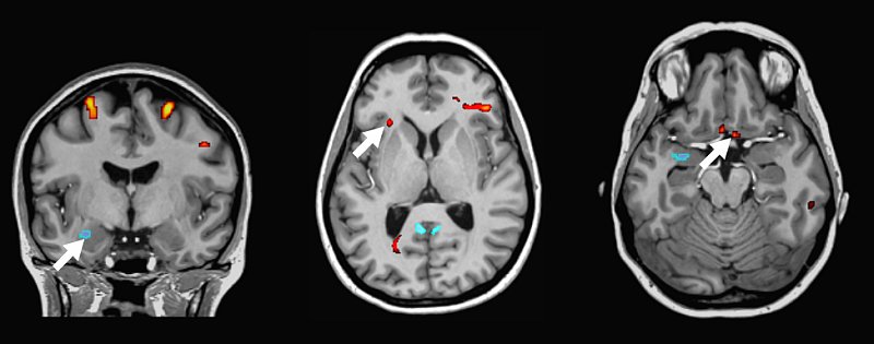
RESEARCH SPOTLIGHT Using MRI to acquire fast, repeatable brain conductivity measurements
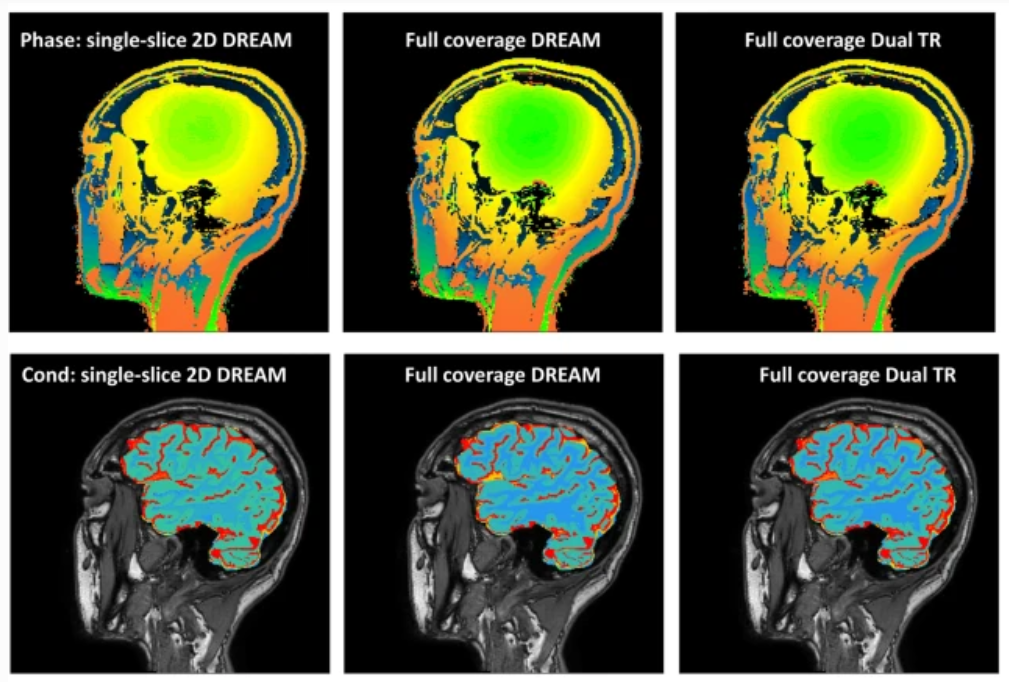
Recent advancements in an imaging technique that measures tissue conductivity and permittivity have positioned it to be a potential candidate for use in many future MRI research studies. Implementation of MREPT in the clinic requires repeatable measurements at a short scan time and an appropriate protocol. Here, we investigated the repeatability of conductivity measurements using …
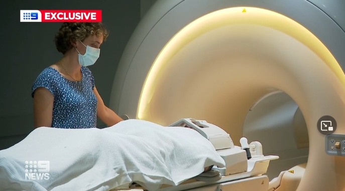
Channel 9 News features imaging trial to treat chronic pain with cannabidiol
A world-first trial conducted at NeuRA Imaging looking at the pain mitigating effects of using cannabidiol has been featured in Chanel 9 News. Spearheaded by Luke Henderson and coordinated by Rebecca Robertson, the trial accesses the NeuRA Imaging Centre to acquire images on spinal cord injury participants. A world-first trial is set to examine whether …
New tool developed at NeuRA for assessing research quality in biomedical publications
The QuOCCA (Quality Output Checklist and Content Assessment) form has been developed by researchers at Neuroscience Research Australia to assist in the evaluation of research quality and reproducibility in biomedical publications. As outlined in a summary at DORA, the 11-item checklist aims to assess research transparency, design and analysis, and reporting practices and can be …
RESEARCH SPOTLIGHT New trial using cannabis derivative for chronic pain following spinal cord injury
Half of all people with spinal cord injury will develop chronic pain—just one of the unseen effects that can be debilitating for the person with an injury. A specific type of pain — neuropathic pain — can be so severe that many regard the pain as the most debilitating consequence of their injury. Neuropathic pain …
Recent Comments